
By Warren R. Heymann, MD
March 11, 2020
Vol. 2, No. 10

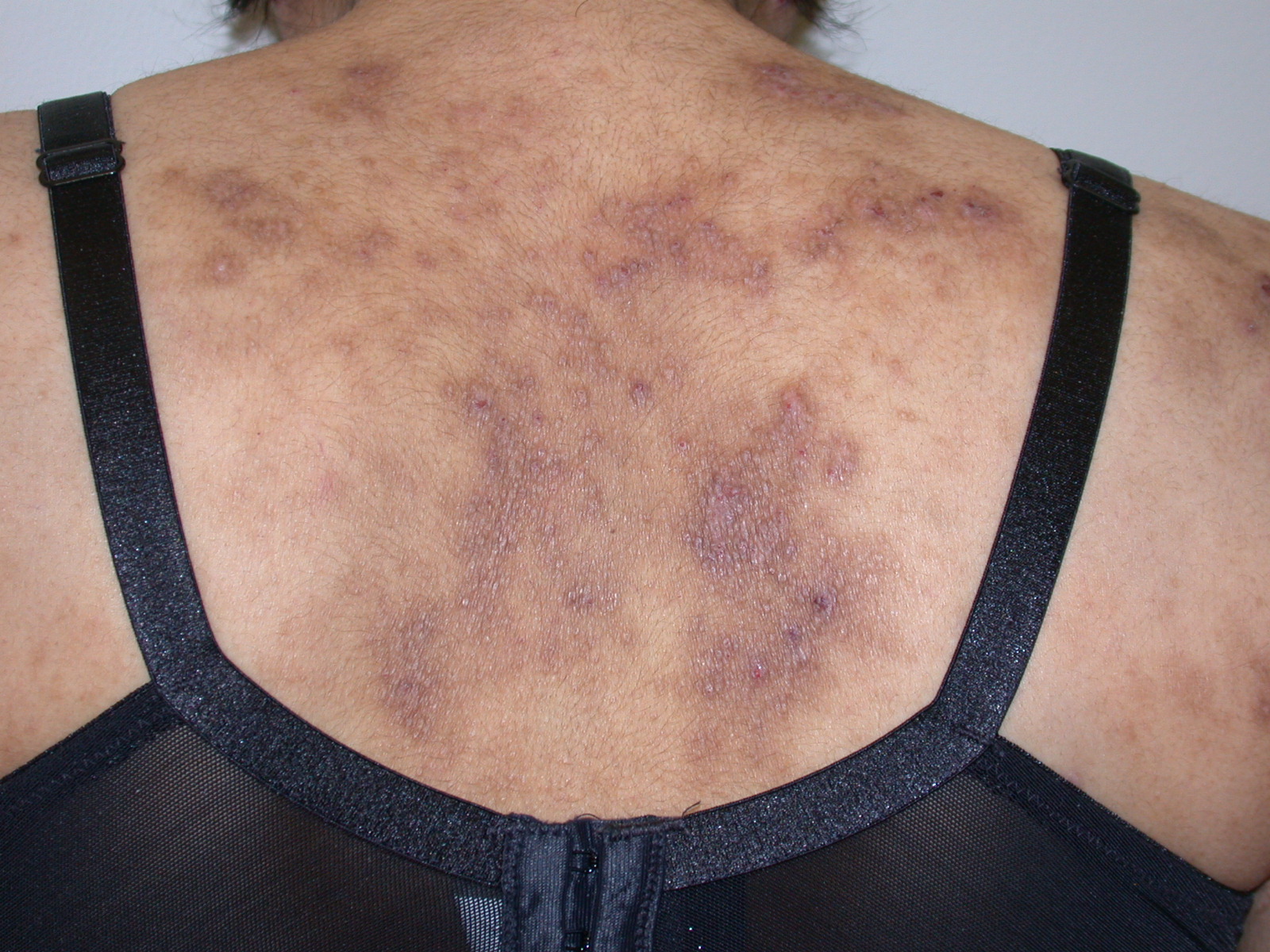
Over the past decade, I have noticed an inversion of the ratio of patients with severe psoriasis versus atopic dermatitis (AD) in my practice. The biologic revolution has clearly favored those afflicted with psoriasis; although dupilumab has improved the lives of many patients suffering from AD, biologic therapy for the disorder is in its relative infancy.
In their outstanding review of advances in AD, Renert-Yuval and Guttman-Yassky note that AD has a “complex, heterogeneous molecular fingerprint among different age groups and ethnicities.” The onset of acute AD skin lesions is characterized by large increases in TH 2/TH 22-related cytokines and chemokines with some TH 17-related signals. Subsequently, intensification of these axes as well as TH 1 augmentation orchestrates the chronic phenotype. (1) Based on these findings, new topical agents, such as JAK (Janus kinase) inhibitors (notably ruxolitinib) and the aryl hydrocarbon receptor modulating agent tapinarof appear promising. Systemic agents, including the oral JAK inhibitors, histamine 4 receptor antagonists, the IL-13 antagonists lebrikizumab and tralokinumab, and the IL-31RA antagonist nemolizumab are on the horizon for advancing the therapeutic armamentarium for AD. (1,2) This commentary focuses on nemolizumab.
According to Mohammed et al, IL-31 is primarily secreted by T-cells, eosinophils, dendritic cells, and macrophages. IL-31 receptors are expressed constitutively on the surface of keratinocytes, eosinophils, and small diameter neurons. The IL-31 receptor complex composed of two subunits, IL-31 receptor alpha (IL-31RA) and oncostatin M receptor beta. Overexpression of interleukin-31, independent of mast cells and lymphocytes, induces clinical and histological features consistent with AD. Activation of IL-31 receptor, directly or indirectly, results in signaling through at least four pathways: JAK-STAT, NF-kB, MAPK, and AKT/PI3K in target cells. In neurons, IL-31-elicited activation induces pruritus and selectively promotes neural growth of small-diameter neurons, activating many of the same genes as nerve growth factor. (3)
Nemolizumab is a monoclonal antibody that antagonizes IL-31RA, thereby blocking IL-31 signaling on its effector cells, including peripheral neurons. An initial randomized, double-blind placebo-controlled phase I/Ib study of nemolizumab demonstrated that a single subcutaneous injection of the drug was well-tolerated in healthy volunteers and patients with AD. It decreased pruritus (approximately 50% based on a pruritus visual analogue scale), sleep disturbance, and topical use of hydrocortisone suggesting that the drug may be a novel therapeutic option for AD. (4)
Ruzicka et al performed a phase II, randomized, double-blind, placebo-controlled, 12-week trial utilizing nemolizumab, assigning adults with moderate-to-severe atopic dermatitis that was inadequately controlled by topical treatments to receive subcutaneous nemolizumab (at a dose of 0.1 mg, 0.5 mg, or 2.0 mg/kg of body weight) or placebo every 4 weeks or an exploratory dose of 2.0 mg/kg of nemolizumab per kilogram every 8 weeks. In total, 216 (82%) completed the study. At week 12, among the patients who received nemolizumab every 4 weeks, changes on the pruritus visual-analogue scale were -43.7% in the 0.1-mg group, -59.8% in the 0.5-mg group, and -63.1% in the 2.0-mg group, versus -20.9% in the placebo group (P<0.01 for all comparisons). Changes on the EASI were -23.0%, -42.3%, and -40.9%, respectively, in the nemolizumab groups, versus -26.6% in the placebo group. Respective changes in body-surface area affected by AD were -7.5%, -20.0%, and -19.4% with nemolizumab, versus -15.7% with placebo. Among the patients receiving nemolizumab every 4 weeks, treatment discontinuations occurred in 9 of 53 patients (17%) in the 0.1-mg group, in 9 of 54 (17%) in the 0.5-mg group, and in 7 of 52 (13%) in the 2.0-mg group, versus in 9 of 53 (17%) in the placebo group. Exacerbation of AD, nasopharyngitis, upper respiratory tract infection, peripheral edema, and increased creatine kinase levels were the most common adverse events in this study. Exacerbation of AD and peripheral edema were more common in the nemolizumab groups than in the placebo group. There were no significant differences observed between the nemolizumab groups and the placebo group regarding creatine kinase. The authors concluded that nemolizumab (at all monthly doses) significantly improved pruritus in patients with moderate-to-severe atopic dermatitis. (5) In follow-up studies, it was demonstrated that nemolizumab was efficacious and well-tolerated for moderate-to-severe AD up to 64 weeks, with patients demonstrating improved activity and productivity at work. (6,7)
Further trials will be necessary to determine if nemolizumab is a viable treatment for AD, either as monotherapy or in combination with other modalities. Inevitably, there will be studies exploring nemolizumab’s role in other pruritic diseases, including those frustrating patients whose recalcitrant pruritus is idiopathic. I will conclude with a quote from Ron Howard: “I think it’s in our nature to try to get beyond that next horizon. I think that when we, as a species, are scratching that itch, we’re actually following an evolutionary compulsion that is wired into us. I think good things come of it.” Time will judge whether nemolizumab is on the horizon in 2020 (or beyond) for AD. Regardless, good things will come of it — whether it is nemolizumab or another inhibitor of the itch cytokine IL-31.
Point to Remember: IL-31 is important as a pruritus-inducing cytokine. Inhibiting the IL-31 receptor by the monoclonal antibody nemolizumab appears to significantly improve AD-associated pruritus.
Our Expert’s Viewpoint
Brian S. Kim, MD, MTR, FAAD
Associate Professor of Medicine (Dermatology)
Co-Director, Center for the Study of Itch
Division of Dermatology, Department of Medicine
Washington University School of Medicine
Originally discovered in 2004, IL-31 was the first cytokine identified to act as an itch-inducing molecule or pruritogen. At the time, it was shown that simply overexpressing IL-31 in mice would lead to atopic dermatitis (AD)-like disease and, most strikingly, itch. Numerous studies followed demonstrating the role of IL-31 in stimulating sensory neurons to evoke itch and its distinct physiology in human AD. Now, in 2019, phase 2 clinical trials have demonstrated anti-itch efficacy of the anti-IL-31 receptor monoclonal antibody nemolizumab in humans with AD and phase 3 clinical trials are currently underway. Importantly, IL-31 biology emboldened the discovery of the role of many other cytokines such as IL-4, IL-13, IL-33, and TSLP in acting in a similar manner. Indeed, agents targeting these pathways including dupilumab, lebrikizumab, tralokinumab, and tezepelumab, respectively, are either on the market or in clinical development.
Dr. Kim had disclosed financial relationships with the following to the AAD at the time of publication: AbbVie, Cara Therapeutics, Incyte Corporation, Kiniksa Pharmaceuticals, Ltd., Menlo Therapeutics, Nuogen Pharma, Pfizer Inc., and Regeneron. Full disclosure information is available at coi.aad.org.
- Renert-Yuval Y, Guttman-Yassky E. What’s new in atopic dermatitis. Dermatol Clin 2019; 37: 205-213.
- Nguyen HL, Anderson KR. Tollefson MM. New and emerging therapies for pediatric atopic dermatitis. Paediatr Drugs 2019; 21: 239-260.
- Saleem M, Oussedik E, D’Amber V, Feldman SR. Interleukin-31 pathway and its role in atopic dermatitis: A systematic review. J Dermatolog Treat 2017; 28: 591-599.
- Nemoto O, Furue M, Nakagawa H, Shiramoto M, et al. The first trial of CIM331, a humanized antihuman interleukin-31 receptor A antibody, in healthy volunteers and patients with atopic dermatitis to evaluate safety, tolerability and pharmacokinetics of a single dose in a randomized, double-blind, placebo-controlled study. Br J Dermatol 2016; 174: 296-304.
- Ruzicka T, Hanifin JM, Furue M, Pulka G, et al. Anti-Interleukin-31 receptor A antibody for atopic dermatitis. N Engl J Med 2017; 376: 826-835.
- Kabashima K, Furue M, Hanifin JM, Pulka G, et al. Nemolizumab in patients with moderate-to-severe atopic dermatitis: Randomized, phase II, long-term extension study. J Allergy Clin Immunol 2018;142(4):1121-1130.
- Mihara R, Kabashima K, Furue M, Nakano M, Ruzicka T. Nemolizumab in moderate to severe atopic dermatitis.: An exploratory analysis of work productivity and activity impairment in a randomized phase II study. J Dermatol 2019; 46: 662-671.